Brief History of nano-scale Imaging and Surface Analysis at the Nanofab
Creating a Facility Housing the Instruments,
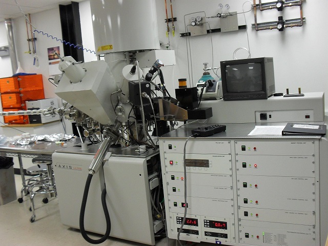
In 2005 Dr. Loren Rieth spearheaded the writing of a successful NSF-CRIF (Chemical Research Instrumentation Fund) PI’ed by distinguished faculty member Scott Anderson of Chemistry. and strongly supported by interim Engineering Dean and Nanofab Director, Robert Roemer. Co-PI’s were Jerry Stringfellow and Ian R. Harvey. The proposal was founded in a strong basis of need from existing research grants, and exhibited a commitment from the University in the form of a dedicated PhD to run the instrument and support analysis interpretation for faculty and student researchers. The grant was awarded on the first submission, and the new Kratos instrument formed the basis of the new Surface Analysis and nano Imaging lab that began to gel around it, in MEB 1555B.
First to respond was Prof Steve Blair of ECE who had been awarded funding to purchase an ellipsometer. But he felt the ellipsometer was better utilized if placed in a common space where many people could use it, and where it could be maintained and supported through training and mentoring by expert, dedicated staff. Dr. Loren Rieth became the first University Surface Scientist, as defined in the NSF CRIF proposal, and he supported the initial applications coming into the lab, and teamed with Prof. Anderson to teach a lecture/lab combo course in Surface Analysis. Soon other complementary instruments were obtained through a variety of means: Zygo non-contacting profilometer (VP instrumentation fund with Zygo cost-share), Polyvar Metallurgical microscope (internal funds), AFM (transfer from faculty Jerry Stringfellow followed by upgrades with internal funds), FE-ESEM/EDS (USTAR and VP funds), XRF and EBSD (donation from benefactors George and Linda Caine). Finally, owing to a favorable trial-run arrangement with FEI, we were able to bring in a quanta dual-beam focused ion beam microscope, which is a FIB with training wheels. It took about 18 months, but we were able to build up the clientele and demand that justified the expenditure of USTAR/MRSEC, VP and faculty USTAR (Solzbacher, Misra) funds to acquire the state-of-the-art dbFIB from FEI, the Helios 650i with five gas injectors and a robotic manipulator for TEM sample prep.
The USTAR Building
In January of 2012, just as the doors were opened to the new Sorenson Molecular Biotechnology (USTAR) Building, the surface lab instruments were the first to move in to the suite named for the first enabling donor of the USTAR building, Micron Technology Inc. Foundation. These first instruments included Prof. Misra’s HR-SEM, quickly followed by the brand new dual-beam focused ion beam instrument. Then the space quickly filled with sample prep tools, the supporting tools mentioned above from the MEB Surface Analysis and nano Imaging lab. It is of interest to note that the Micron Technology Inc. Foundation Suite was designed to support many instruments, but uniquely (with perhaps three peer campuses) these instrument rooms were envisioned in a way that included both life science imaging instruments and hard materials (physical sciences) imaging instruments, all in one space. However at the time of programming, it was never anticipated exactly how those spaces would be outfitted with the expensive instruments. In a case study of “build it and they will come”, the space allocated for confocal microscopy was soon filled when Prof. Ghandehari offered his Olympus FV-1000 to become available for open access by others, and for care and support by the Nanofab staff. In 2013 the Health Sciences Center (HSC cores Director Dr. John Philips and EM Core director Dr. David Belnap) enquired about relocating their EM core to the same suite, and it did not take long to bring that to fruition, filling space that was exactly envisioned for that function. Two Bio TEM’s and associated sample preparation with Dr. Belnap and five technicians relocated to the space in 2013. Working with the MRSEC team and USTAR funds, we were able to finally bring analytical S/TEM back to the University after nearly a 15 year hiatus. JEOL created an extremely favorable partnership with the University to bring in their new first-of-its-kind atomic imaging and ultrafast 3D elemental tomography instrument, and make it the north American demo site for that instrument. Through all of this growth, we are also pleased to acknowledge the sequential addition of expert professional scientific support of Dr. Brian Van Devener (physical/analytical/surface chemistry), Dr. Randy Polson (laser optics and high resolution electron and ion microscopy), and Dr. Paulo Perez (physical chemistry and high resolution scanning and transmission electron microscopy). At the beginning of 2014, the circle was closed again, with the addition of Distinguished Professor of Chemistry Scott Anderson joining as the Associate Director for the facility, functioning as the Associate Director and Faculty Advocate.
In 2015 we took delivery of our JEOL 2800 analytical STEM with fast EDS, which was part of the USTAR cost-share to the NSF/MRSEC grant. This instrument boasts dual EDS detectors with ultrafast collection, making possible high resolution EDS mapping in 2D and 3D. Thanks to Prof. Siva Guruswamy for spearheading this acquisition. In 2017 we installed two major upgrades to the S/TEM: Protochips liquid phase and gas phase in-situ experimentation. In this case, Prof. Scott Anderson wrote two grants (one intramural and one DURIP) to enable the upgrades. It was a good year: Prof. Stephen Naleway brought in a Hysitron nanoindenter and Prof. Ashley Spear brought in EDS/EBSD for the FIB instrument.