Interactive Equipment Map


 |
fullname test test description |
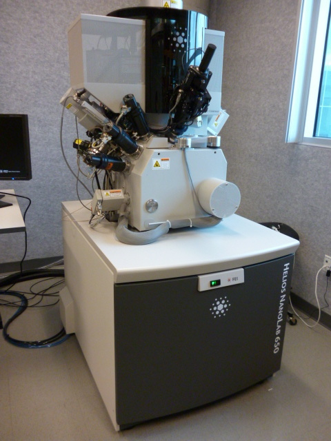 |
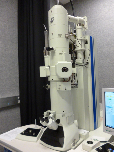
dbFIB, FEI Helios NanoLab 650 A few key capabilities of the instrument include: Slice-n-view: the ability to incrementally image then cut 10 nm slices into the face of a 5um X 5um area, then rebuilding the images into a tomographic volume. Deposit Pt in pre-defined patterns, down to ~35 nm line widths. Mill patterns using the FIB as a precision scalpel. Create precision cross-sections for in-situ viewing. Selectively remove organic material by precision FIB cutting, using injected water vapor enhanced etching. UHR- SEM column: with sub nm resolution. Omniprobe tool, gives the ability to prepare samples for TEM analysis |
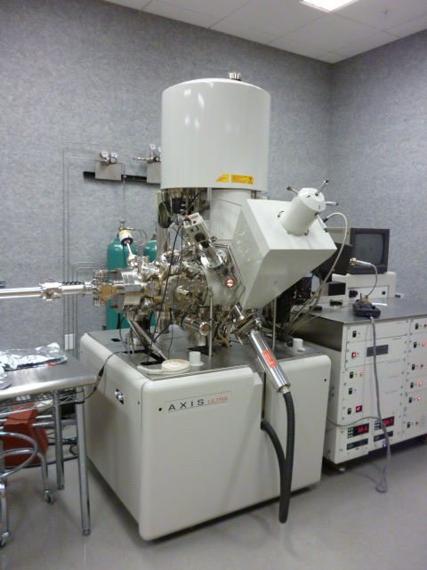 |
XPS, Kratos Axis Ultra DLD This system is a state-of-the-art multitechnique surface analysis tool. The system measures the chemical composition and bonding of a surface, and can also perform depth profiles which look at the composition below the surface to limited depths. The system has been acquired through generous funding from an NSF Chemistry Research Instrumentation grant (CHE-0443657) and matching funds from the University of Utah Office of the Vice-president for Research. |
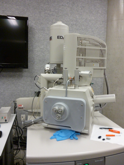 |
SEM, FEI Quanta 600 FEG The Quanta 600 is a State-of-the-Art scanning electron microscope (SEM) with a high-resolution field-emission source. The system can operate in high-vacuum, low-vacuum, and true environmental (water vapor ambient) modes. The electron detectors included with this system are an Everhart-Thornley secondary electron detector, a large-field detector, a gaseous secondary electron detector, and a solid-state backscattered electron detector. A peltier cooled sample holder is available to aid in imaging biological and "wet" specimens. Analytical options will include an EDX system for compositional analysis, and an electron backscattered diffraction (EBSD) system for phase ID and to measure crystal orientation. This instrument was purchased through exceptionally generous support from the Office of the Vice-President for Research, with the EBSD and complementary XRF system provided through the generous donation of George and Linda Caine. |
| |
fullname test description |
| |
fullname test description |
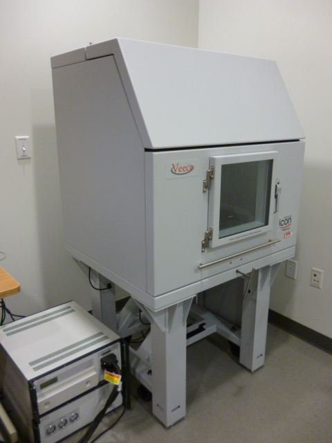 |
AFM, Bruker Dimension Icon We are pleased to announce that we have just acquired a new state-of-the-art Dimension ICON-PT atomic force microscope (AFM). Three major new capabilities of this system relative to the old system are a closed-loop scanner, ScanAsyst, and PeakForce QNM. These new capabilities increase the precision for our measurements, decrease user intervention necessary to set gains, and allow routine and high-resolution mapping of the mechanical properties of films, respectively. This system replaces our Dimension 3000, and continues to offer contact and tapping modes. |
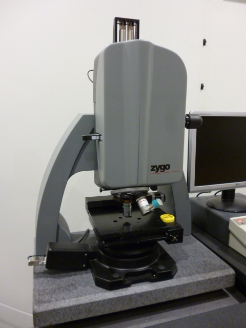 |
Optical Profiler, Zygo NewView Acquired through University Vice-President support in conjunction with a major contribution from the Zygo Corporation, this instrument quantitatively measures surface topography of a sample using white light optical interference. This yields a non-contact and very rapid method to acquire 3-D surface data over the field-of-view for the objective lens used. Common applications include surface roughness quantification, MEMS inspection, MEMS device characterization, and etch inspection. |
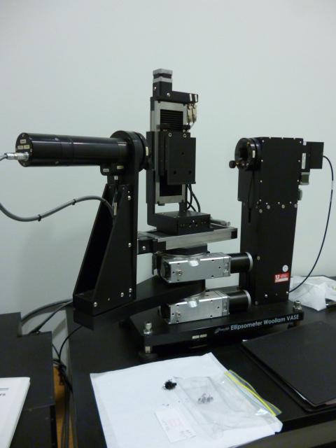 |
Ellipsometer, Woollam Spectroscopic This instrument is a Variable Angle Spectroscopic Ellipsometer (VASE) which has small spot focusing optics and an automated X-Y translation stage. This instrument is primarily used to measure the thickness and optical properties (index of refraction and extinction coefficient) of thin film materials. These measurements are made by analyzing polarized light reflected from the sample surface, and fitting the measured data to a model. Typically dielectric and semiconductor films are measured, though very thin metal films can also be analyzed. |
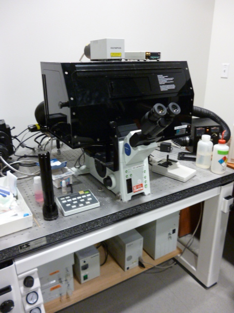 |
Confocal Microscope, Olympus FV-1000 This laser scanning confocal microscope has a variety of excitation laser sources and three separate detector channels. The motorized stage allows sample acquisition from many locations. The computer controlled filter selections means the user simple selects the appropriate dyes from a list and the instrument then configures the laser excitation sources and detector channels. |
 |
fullname test description |
 |
fullname test description |
 |
Bruker D8 Discover X-Ray Diffractometer (XRD). The system offers thorough non-destructive crystal phase characterization of thin films via X-ray reflectometry (XRR), high resolution x-ray diffraction (HRXRD), reciprocal space mapping (RSM) and grazing incidence diffraction (GID). A vacuum attachment allows the instrument's sample holder to map and analyze 12" and 6" wafers. |
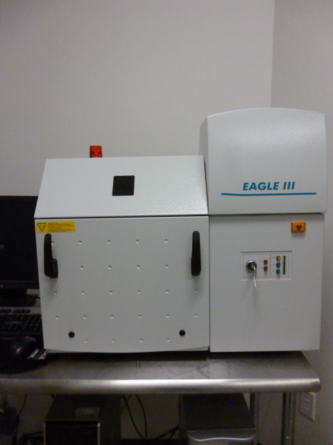 |
XRF, Eagle III Microspot EDS-based X-ray fluorescence (XRF) provides sub 1% bulk compositional analysis of large samples. The scanning stage enables elemental mapping (referenced to an optical image), with 0.1mm resolution, using microfocus capillaries. Made possible by a generous donation from George and Linda Caine. |
 |
fullname test description |
 |
fullname test description |
 |
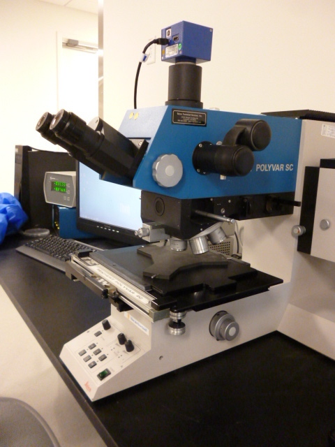
Microscope, Polyvar MET Optical microscopy is a great first step for characterizing surfaces. The scope has bright-field, dark-field, and Nomarski (Differential Interference Contrast) imaging modes, and a 5 MP digital camera to acquire publication quality images. Optical microscopy is an excellent way to become familiar with your sample surface before using more exotic and potentially artifacted measurement tools. |
 |
fullname test description |
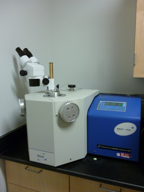 |
Fischione Ion mill Model 1060 Ion milling is used to enhance the quality of a sample's surface. Inert gas, typically argon, is ionized and then accelerated toward the sample. By means of momentum transfer, the impinging ions sputter material at a controlled rate. Two TrueFocus ion sources direct controlled-diameter ion beams at variable angles to the sample to allow rapid milling at high beam energy and accurate polishing at low energy. |